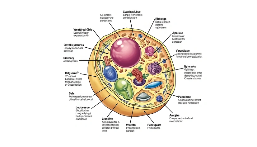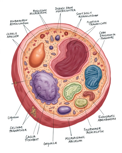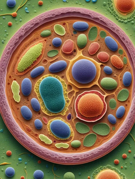
animal cell diagram labeled
Resize and add details
Convert to video
Outpaint the rest of the image
Additional Info
Steps7
SamplerK_EULER_ANCESTRAL
CFG Scale2
Seed783458431
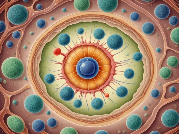
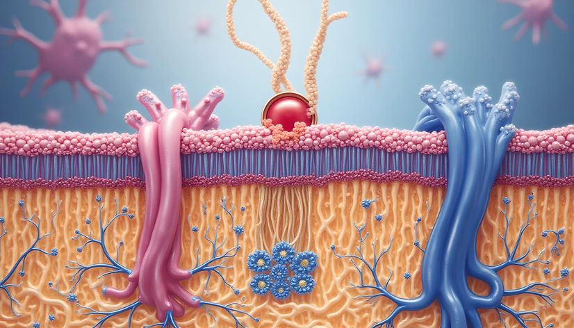
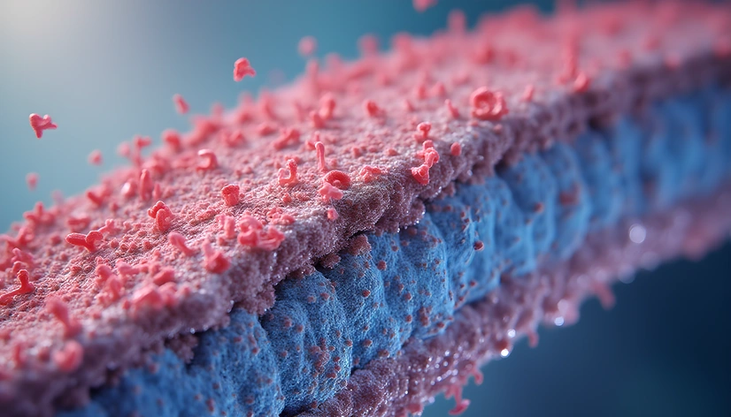
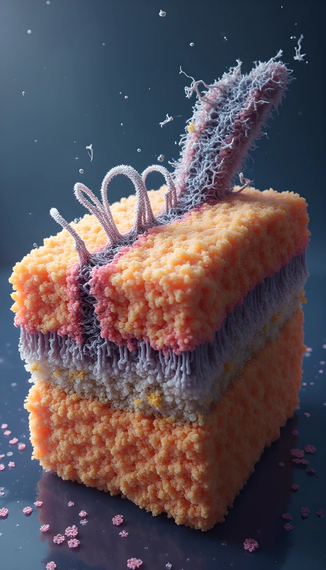
Enhanced Promptcentrioles, paired cylindrical organelles involved in microtubule formation, situated near nucleus, labeled in dark blue, mitochondria, double-membraned organelles responsible for cellular respiration, generating energy through ATP production, depicted in bright red, rough endoplasmic reticulum, extensive network of membranous tubules and flattened sacs, studded with ribosomes, facilitating protein synthesis and transport, shown in pale blue, smooth endoplasmic reticulum, tubular structure involved in lipid synthesis and detoxification, lacking ribosomes, displayed in light green, Golgi apparatus, complex organelle consisting of flattened sacs and tubules, responsible for protein modification and packaging, illustrated in vibrant orange, lysosomes, membrane-bound vesicles containing digestive enzymes, breaking down and recycling cellular waste, represented in bright yellow, nucleus, membrane-bound organelle containing genetic material, regulating cellular activities, portrayed in deep purple, nucleolus, region within nucleus responsible for ribosome synthesis, visible in pale gray, plasma membrane, thin semi-permeable lipid bilayer, surrounding cell and controlling movement of substances, depicted in dark gray, ribosomes, small organelles found throughout cytoplasm, responsible for protein synthesis, shown in bright pink, cytoplasm, jelly-like substance filling cell, containing various organelles and structures, displayed in pale green, cytoskeleton, network of protein filaments providing structural support and shape to cell, illustrated in dark brown, microvilli, small projections on cell surface, increasing surface area for absorption and interaction, represented in light brown, cell wall, rigid outer layer providing support and protection, visible in dark green, chloroplasts, organelles found in plant cells, responsible for photosynthesis, displayed in bright green, large central vacuole, storage organelle containing water, salts, and waste products, portrayed in pale blue, peroxisomes, small organelles involved in fatty acid breakdown and detoxification, represented in bright orange, endosomes, membrane-bound vesicles involved in cellular transport and sorting, shown in light gray, phagosomes, membrane-bound vesicles containing ingested particles, illustrated in dark gray, autophagosomes, double-membraned vesicles involved in cellular recycling and degradation, depicted in pale yellow, exosomes, small extracellular vesicles involved in intercellular communication, represented in light blue, gap junctions, specialized channels connecting adjacent cells, facilitating direct communication, shown in bright blue, tight junctions, specialized structures sealing adjacent cells together, regulating permeability, illustrated in dark green, adherens junctions, specialized structures connecting adjacent cells, maintaining tissue structure, portrayed in pale green, desmosomes, specialized structures connecting adjacent cells, providing mechanical strength, represented in bright red, hemidesmosomes, specialized structures connecting cells to basement membrane, maintaining tissue structure, depicted in dark red, synapses, specialized structures connecting neurons, facilitating chemical transmission, shown in bright yellow, dendrites, branching extensions of neurons, receiving and processing signals, illustrated in pale blue, axons, long extensions of neurons, transmitting signals, portrayed in dark blue, myelin sheath, insulating layer surrounding axons, facilitating signal transmission, represented in bright white, nodes of Ranvier, gaps in myelin sheath, facilitating signal transmission, depicted in pale gray, axon terminals, specialized structures releasing neurotransmitters, shown in bright pink, postsynaptic density, specialized structure receiving and processing signals, illustrated in dark gray, synaptic cleft, small gap between neurons, facilitating chemical transmission, portrayed in pale green.