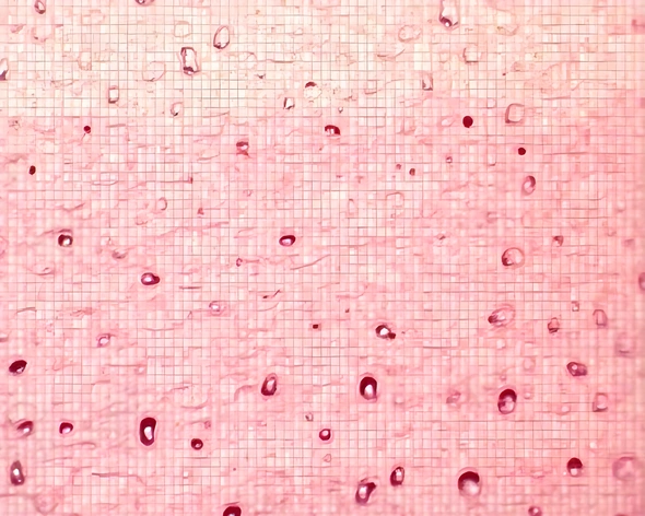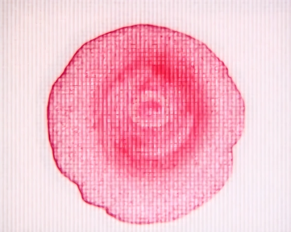
connective tissue under microscope
Resize and add details
Convert to video
Outpaint the rest of the image
Additional Info
ModelFlux
Seed640022321
Enhanced Promptcellular structure, connective tissue, dense collagen fibers, cells embedded within a gel-like matrix, blood vessels and nerves running alongside, fibroblasts producing new collagen, ground substance composed of glycoproteins and proteoglycans, hyaluronic acid, cellular morphology varying between loose and dense arrangements, histological staining techniques used to highlight specific components, transmission electron microscopy providing ultrastructural details, scanning electron microscopy showing surface topography, confocal laser scanning microscopy capturing three-dimensional views, fluorescent labeling techniques applied to visualize specific cell types, fluorescence microscopy revealing cell-cell interactions, phase contrast microscopy highlighting refractive index differences, DIC microscopy differentiating between living and dead cells, brightfield microscopy contrasting nuclear and cytoplasmic features, hematoxylin and eosin stain used for general histology, Masson's trichrome stain distinguishing collagen and muscle fibers, Gomori's methenamine silver stain highlighting elastic fibers, Weigert's iron-hematoxylin-safranin stain detecting reticular fibers, transmitted polarized light demonstrating birefringence of collagen, immunohistochemistry identifying specific proteins and antigens, molecular biology techniques analyzing gene expression and protein function, fluorescence in situ hybridization localizing mRNA and chromosomal regions, super-resolution microscopy resolving subcellular structures, nanoscale imaging techniques visualizing extracellular matrix components, advanced computational models simulating tissue mechanics and dynamics, interdisciplinary approaches integrating biology, physics, and mathematics, high-throughput screening technologies identifying novel therapeutic targets, regenerative medicine strategies exploiting tissue engineering principles, tissue clearing and expansion methods preserving structural integrity, multiphoton microscopy probing deeper tissues without photobleaching, photoacoustic microscopy combining optical and ultrasound imaging, photo-induced force microscopy measuring forces at the nanoscale, atomic force microscopy characterizing surface topography and elasticity, Raman spectroscopy identifying molecular composition and orientation, infrared spectroscopy detecting vibrational modes of molecules, x-ray scattering and diffraction techniques determining crystallographic structures, synchrotron-based imaging providing high-energy radiation sources, cryo-electron tomography reconstructing three-dimensional images, correlative microscopy combining multiple modalities for comprehensive analysis, super-resolution localization microscopy resolving individual biomolecules, single-molecule localization microscopy tracking dynamic processes at the nanoscale, stimulated emission depletion (STED) microscopy reducing point spread function, structured illumination microscopy improving resolution and sensitivity, total internal reflection fluorescence microscopy detecting near-surface phenomena, coherent anti-Stokes Raman scattering microscopy enabling label-free detection, optical coherence tomography imaging subsurface structures non-invasively, multiphoton microscopy penetrating deeper tissues without photobleaching, nonlinear optical microscopy generating high-contrast images, Fourier transform infrared spectroscopy analyzing molecular vibrations, second harmonic generation microscopy detecting nonlinear optical responses, third harmonic generation microscopy producing high-contrast images, two-photon excitation microscopy exciting fluorophores with reduced photobleaching, two-color fluorescence microscopy separating overlapping spectral signals, time-resolved fluorescence microscopy monitoring dynamic processes, Förster resonance energy transfer microscopy transferring energy