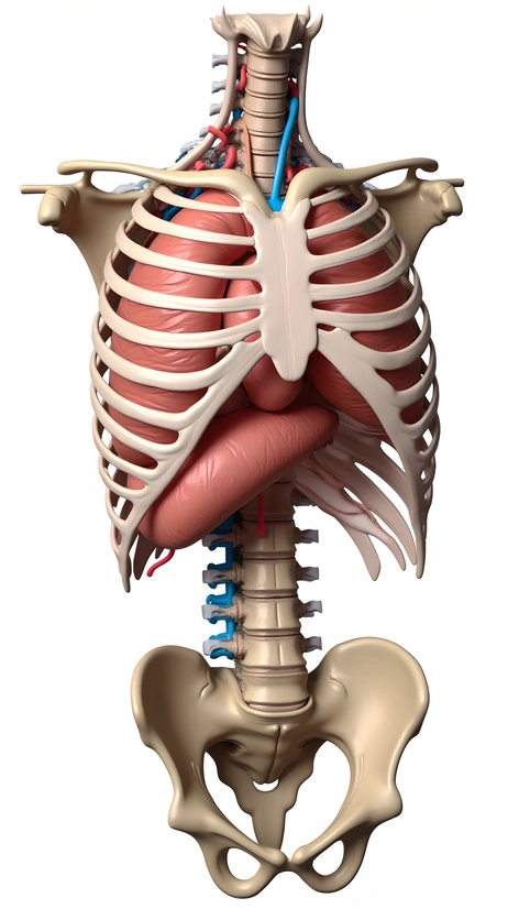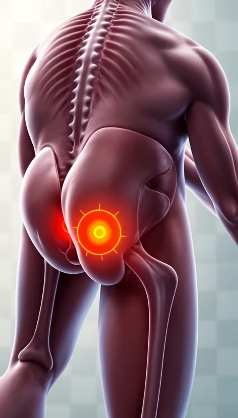
hip pain location diagram
Resize and add details
Convert to video
Outpaint the rest of the image
Additional Info
Steps28
Samplerddim
CFG Scale8.3
Seed960147092
Enhanced PromptDetailed diagram of hip pain locations, anterior view, posterior view, labeled illustrations, pain zones highlighted, color-coded for varying levels of discomfort, anatomical structures clearly visible, bones, muscles, tendons, ligaments, nerves, femur, pelvis, sacrum, coccyx, piriformis muscle, sciatic nerve, iliotibial tract, hip joint, joint capsule, acetabulum, femoral head, femoral neck, greater trochanter, lesser trochanter, medical and anatomical accuracy, precise labeling, arrows indicating pain radiating to surrounding areas, including lower back, buttocks, thigh, knee, leg, foot, visual representation of pain patterns, assisting diagnosis and treatment of hip-related issues, such as arthritis, bursitis, tendinitis, strains, and sprains, educational tool for healthcare professionals, patients, and students, enhancing understanding of hip anatomy and pain management.