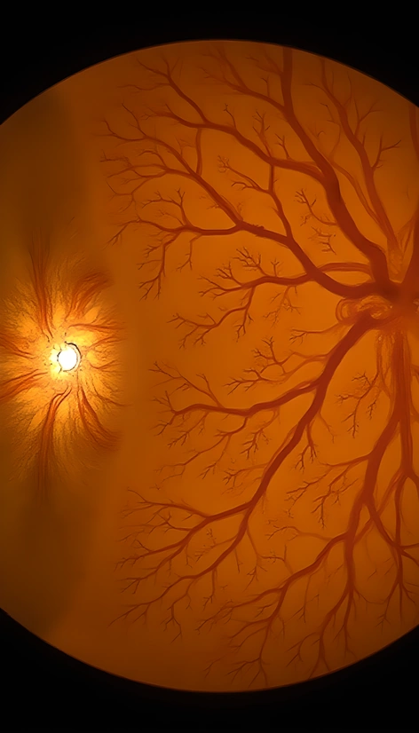
photography retinal fundus images
Resize and add details
Convert to video
Outpaint the rest of the image
Additional Info
ModelFlux
Seed456803011
Enhanced Prompthigh-resolution, high-quality retinal fundus images, ophthalmology photography, posterior segment imaging, detailed vascular pattern visualization, clear visibility of optic disc, macula, and fovea, subtle color gradations, precise control over illumination, balanced contrast, accurate representation of anatomical structures, minimal artifacts, no motion blur, sufficient magnification, well-defined borders, soft focus effect on surrounding ocular tissues, clinical-grade equipment, standardization of image acquisition parameters, consistent color calibration, strict adherence to medical imaging protocols, DICOM-compliant file format, spatial resolution of at least 1024x768 pixels, 8-bit or 16-bit color depth, acquired using a high-end fundus camera, preferably with adaptive optics technology, patient eye aligned precisely with the camera axis, proper fixation target used, image captured during a single blink-free interval, ambient light controlled, no glare or reflections on the cornea, careful attention to patient positioning and comfort, strict hygiene and infection control practices followed, images stored securely and confidentially in accordance with HIPAA regulations, anonymized data available upon request, consent obtained from participants or their guardians, study design adheres to IRB-approved guidelines, results published in a peer-reviewed journal, data shared openly on a publicly accessible database, metadata included for reproducibility and transparency