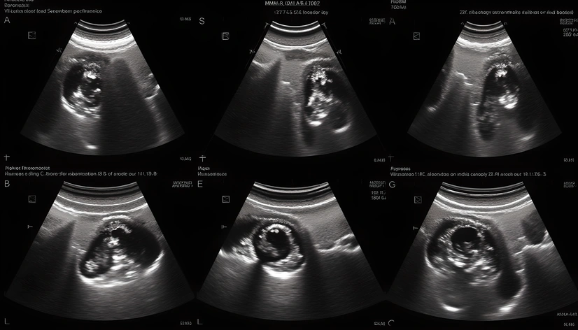
thyroid ultrasound photos
Resize and add details
Convert to video
Outpaint the rest of the image
Additional Info
ModelFlux
Seed1000952957
Enhanced Promptthyroid ultrasound images, grayscale mode, transverse section, sagittal section, longitudinal section, thyroid gland clearly visible, surrounding tissues distinguishable, clear delineation between thyroid tissue and adjacent structures, prominent nodule or cyst highlighted, calcifications marked, vascular structure labeled, lymph nodes identified, measurement of thyroid nodule size and location, clear labeling of key anatomical features, standard ultrasound machine settings, high-resolution probe used, optimal patient positioning for clear imaging, anterior neck region scanned, posterior neck region scanned, parotid glands examined, carotid arteries visualized, jugular veins imaged, soft tissue contrast optimized, speckle pattern minimal, artifacts avoided, clear and concise legend included, medical jargon minimized, diagrammatic annotations provided, educational purposes, teaching tool for students, clinical reference for professionals, peer-reviewed publication quality, precise measurements reported, data analysis software integrated, DICOM format, HIPAA compliance ensured, patient confidentiality maintained, consent obtained, institutional review board approval secured