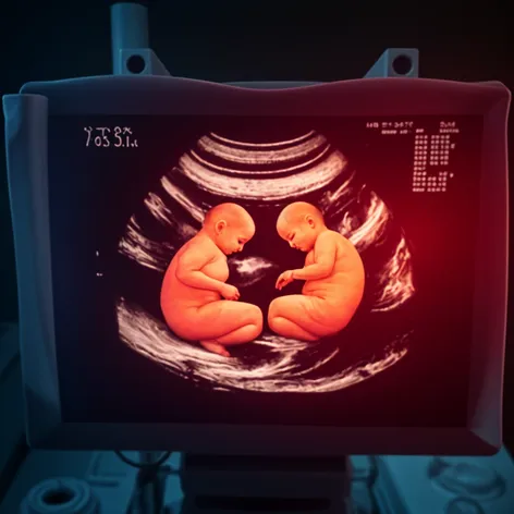
Twin 2 month ultrasound
Resize and add details
Convert to video
Outpaint the rest of the image
Additional Info
Steps25
SamplerDPMSolver++
CFG Scale2
Seed846852454
Enhanced PromptMonochorionic diamniotic twins, 2 months old, ultrasound image, clear visualization of two separate embryos, each within their own amniotic sac, sharing a single placenta, visible yolk sacs, embryonic poles, and fetal membranes, dating ultrasound to confirm gestational age, transabdominal approach, high-frequency probe, 2D grayscale imaging, optimal fetal position for measurement, fetal crown-rump length, biparietal diameter, and abdominal circumference, visible fetal heart activity, normal fetal development, twin-to-twin comparison, assessment of amniotic fluid volume, placental location and morphology, umbilical cord insertion, and membrane thickness, sonographer's annotations and measurements, ultrasound machine settings optimized for fetal imaging, acoustic output and thermal index within safe limits, ultrasound gel and probe placement ensuring optimal image quality, maternal bladder fullness and fetal position optimized for imaging, ultrasound exam performed by a certified sonographer, images reviewed and interpreted by a board-certified radiologist or obstetrician.